- Methodology
- Open access
- Published:
Engineered extracellular vesicles for ischemic stroke: a systematic review and meta-analysis of preclinical studies
Journal of Nanobiotechnology volume 21, Article number: 396 (2023)
Abstract
Background
This systematic review and meta-analysis aimed to evaluate the efficacy of engineered extracellular vesicles (EEVs) in the treatment of ischemic stroke (IS) in preclinical studies and to compare them with natural extracellular vesicles (EVs). The systematic review provides an up-to-date overview of the current state of the literature on the use of EEVs for IS and informs future research in this area.
Methods
We searched PubMed, EMBASE, Web of Science, Cochrane Library, and Scopus databases for peer-reviewed preclinical studies on the therapeutic effect of EEVs on IS.Databases ranged from the inception to August 1, 2023. The outcome measures included infarct volumes, neurological scores, behavioral scores, apoptosis rates, numbers of neurons, and levels of IL-1β, IL-6, and TNF-α. The CAMARADES checklist was used to assess the quality and bias risks of the studies. All statistical analyses were performed using RevMan 5.4 software.
Results
A total of 28 studies involving 1760 animals met the inclusion criteria. The results of the meta-analysis showed that compared to natural EVs, EEVs reduced infarct volume (percentage: SMD = -2.33, 95% CI: -2.92, -1.73; size: SMD = -2.36, 95% CI: -4.09, -0.63), improved neurological scores (mNSS: SMD = -1.78, 95% CI: -2.39, -1.17; Zea Longa: SMD = -2.75, 95% CI: -3.79, -1.71), promoted behavioral recovery (rotarod test: SMD = 2.50, 95% CI: 1.81, 3.18; grid-walking test: SMD = -3.45, 95% CI: -5.15, -1.75; adhesive removal test: SMD = -2.60, 95% CI: -4.27, -0.93; morris water maze test: SMD = -3.91, 95% CI: -7.03, -0.79), and reduced the release of proinflammatory factors (IL-1β: SMD = -2.02, 95% CI: -2.77, -1.27; IL-6: SMD = -3.01, 95% CI: -4.47, -1.55; TNF-α: SMD = -2.72, 95% CI: -4.30, -1.13), increasing the number of neurons (apoptosis rate: SMD = -2.24, 95% CI: -3.32, -1.16; the number of neurons: SMD = 3.70, 95% CI: 2.44, 4.96). The funnel plots for the two main outcome measures were asymmetric, indicating publication bias. The median score on the CAMARADES checklist was 7 points (IQR: 6–9).
Conclusions
This meta-analysis shows that EEVs are superior to natural EVs for the treatment of IS. However, research in this field is still at an early stage, and more research is needed to fully understand the potential therapeutic mechanism of EEVs and their potential use in the treatment of IS.
PROSPERO registration number
CRD42022368744.
Introduction
Ischemic stroke (IS) is a common neurological disease and a leading cause of disability and death [1, 2]. Despite significant advances in the treatment of IS over the past decade, current treatment options are still limited. Tissue plasminogen activator is the only drug approved by the Food and Drug Administration (FDA) for IS treatment, which has a very narrow treatment window of 4.5 h [3, 4]. Mechanical thrombectomy is also only available to 10% of patients [5]. Therefore, there is an urgent need to develop new therapeutic strategies to improve IS prognosis.
Natural extracellular vesicles (EVs) are small vesicles released by cells, carrying a variety of bioactive molecules, such as proteins, lipids and nucleic acids [6]. EVs include exosomes, microvesicles (MVs) and apoptotic bodies [7]. Existing preclinical studies and clinical trials have demonstrated the potential of EVs to treat IS [8]. The biological advantages of EVs include low immunogenicity, natural stability and high delivery efficiency. In addition, EVs can easily cross the blood-brain barrier (BBB), indicating their availability to target central nervous system diseases [9, 10]. Research shows that EVs can regulate angiogenesis, inhibit inflammation, inhibit neuronal apoptosis, reduce cell remodeling, and improve the microenvironment of post-IS brain injury via delivering bioactive molecules to the brain [11,12,13,14,15]. For example, stem cell-derived EVs have been shown to up-regulate the expression of genes of multiple angiogenesis pathways such as vascular endothelial growth factor (VEGF), angiopoietin 1 (ANG1), angiopoietin 2 (ANG2), matrix metallopeptidase 9 (MMP9), thrombopoietin 1 (TSP1), so as to promote the formation of new blood vessels and improve cerebral blood flow [16]. EVs can also deliver brain-derived neurotrophic factor (BDNF) to the brain and promote the survival and growth of neurons [17].
However, EVs limitations suggest low yield, low biological activity, weak targeting and rapid clearance in vivo [8]. To address the limitations of natural EVs as a treatment option for IS, researchers are developing engineered extracellular vesicles (EEVs) through bioengineering techniques [12, 13]. EEVs can be produced in two ways: by engineering modification on EV-producing parent cells or by directly modifying EVs [12]. The first method involves co-incubating the parent cells with targeted molecules or transfecting plasmid into the parent cells to create EVs with specific targeted molecules [18]. The second method involves direct modification of EVs using techniques such as incubation, ultrasonic treatment, electroporation, antibody-specific loading, freeze-thaw methods, and saponin-assisted method [18].
Although there have been many preclinical studies on the treatment of IS with EEVs, there is still a lack of evidence-based research. Therefore, this systematic review and meta-analysis aimed to compare the efficacy differences between EEVs and natural EVs in preclinical studies, providing a basis for future research in this field.
Methods
This systematic review was conducted in accordance with the Preferred Reporting Items for Systematic Reviews and Meta-Analyses (PRISMA) guidelines [19], and the protocol for this meta-analysis has been published in the PROSPERO database with the registration number CRD42022368744.
Search strategy
We conducted a comprehensive literature review to identify relevant studies published in PubMed, EMBASE, Web of Science, Cochrane Library, and Scopus databases until August 1, 2023. We also manually searched for references in relevant reviews and meta-analyses. We aimed to capture all relevant studies on the therapeutic use of EEVs for IS. Our search strategy, therefore, included the use of MeSH terms and free words “extracellular vesicles” and “stroke,“ and we did not impose any restrictions on the term “engineered.“ Details of our retrieval strategies for each database can be found in the supplementary materials.
Study selection
After removing duplicates, we screened titles and abstracts to exclude clearly irrelevant studies and then read the full text of the remaining studies to screen them further based on inclusion and exclusion criteria. The inclusion criteria included: (a) studies published in English, (b) studies conducted on IS animal models, (c) studies providing detailed methods for extracting, engineering, and identifying EVs, (d) Studies that measure at least one of the following: infarct volume or neurological scores. The exclusion criteria included: (a) studies with data that could not be extracted, (b) studies that do not compare the treatment effects of EEVs versus natural EVs, (c) non-rodent studies. Two authors (RY, SHW) independently performed the study selection, and any discrepancies were resolved through discussion with a third author (QH).
Data extraction
We extracted the following information from the included studies: (a) General Information: First author, year of publication, and country. (b) Animal Characteristics: Species, age, gender, weight, and number. (c) EVs Characteristics: Source, extraction method, diameter, engineered method, and engineering targets. (d) Details of EEVs Treatment: Route, dosage, and time. (e) Duration of Follow-Up. (f) Outcome Measures: Infarct Volume: Percentage of the infarct volume and the size of the infarct volume. Neurological Scores: Modified neurological severity score (mNSS) and Zea-Longa score. Behavioral Recovery: Rotarod test, grid-walking test, adhesive removal test, and Morris water maze test. Pro-inflammatory Factor Release: IL-1β, IL-6, and TNF-α. Cellular Effects: Apoptosis rate and neuron numbers .
For studies with only graphical data available, we used the online tool WebPlotDigitizer (https://automeris.io/WebPlotDigitizer/) to extract data from the graphs. If multiple time points were involved throughout the measurements, we extracted data only from the latest time point. Two authors (YHC, JBC) independently performed data extraction, and any differences of less than 10% were averaged while any differences greater than 10% were discussed and resolved with a third author (LY). If additional information was needed, we contacted the authors via email. We excluded these studies if data were still unavailable after two attempts.
Risk of bias assessment
We assessed the quality of the studies using the Collaborative Approach to Meta-analysis and Review of Animal Data in Experimental Studies (CAMARADES) bias risk checklist [20]. This checklist comprises: (a) publication in a peer-reviewed journal, (b) statement of temperature control, (c) randomization, (d) allocation concealment, (e) blinded outcome assessment, (f) avoidance of clearly biased anesthetics, (g) use of appropriate animal models, (h) sample size calculation, (i) compliance with animal welfare regulations, (j) statement of potential conflicts of interest. Two independent authors (XYL and XZ) conducted a risk assessment and any discrepancies were resolved through discussion with a third author (RY).
Statistical analysis
We used Revman 5.4 software for data analysis. Continuous variables were presented as standardized mean difference (SMD) with 95% confidence intervals (CI). We tested for heterogeneity in each outcome measure using the Q-test and I2 statistic. Based on the heterogeneity, we used a fixed-effects model only when no significant heterogeneity was observed (p > 0.1, I2 < 50%). Otherwise, a random-effects model was employed for meta-analysis, with p < 0.05 considered statistically significant. When significant heterogeneity was present, we conducted sensitivity analysis by excluding individual studies one by one and performed subgroup analysis to determine the source of heterogeneity.
Results
Study characteristics
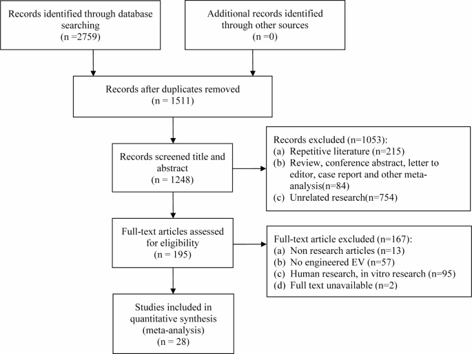
We identified 2793 studies from the databases, which we then screened based on our inclusion and exclusion criteria. As shown in Fig. 1 and 28 studies [17, 21,22,23,24,25,26,27,28,29,30,31,32,33,34,35,36,37,38,39,40,41,42,43,44,45,46,47] ultimately met our criteria and were included in this review. Details of these studies are presented in Table 1. All studies were conducted using rats (n = 19) and mice (n = 9). Apart from two studies that utilized photochemistry and electrocoagulation techniques, the prevalent approach was the suture method of middle cerebral artery occlusion (MCAO) (n = 26). Mesenchymal stem cells (MSC) were the primary source of EVs in most studies (n = 15), with other sources including neural stem cells (NSC) (n = 5), blood (n = 5), and soma (n = 3). The predominant method of engineering EVs was through lentiviral transfection (n = 16), followed by coculture (n = 7), ultrasonic techniques (n = 3), electroporation (n = 1), and surface modification (n = 1). The preferred route of EEVs administration was intravenous injection (n = 21), though some studies opted for intracerebral injection (n = 5) or nasal administration (n = 2). Administration timing varied, spanning from a day before IS (n = 2) to 14 days post-IS (n = 26), with select studies administering EVs on multiple occasions (n = 5). Notably, a significant portion of studies engineered the parent cells (n = 19), as opposed to directly engineering the EVs (n = 9).
Outcomes
EEVs reduce infarct volume and improve neurological scores after IS
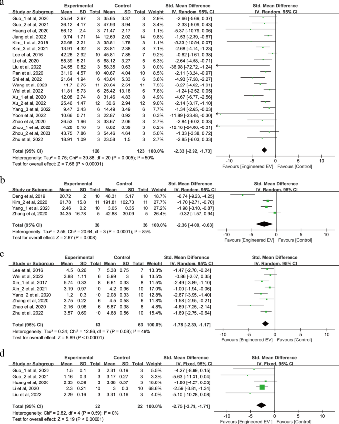
The effects of EEVs therapy on infarct volume and neurological scores were shown in Fig. 2a-d. A total of 321 animals in 25 studies reported changes in infarct volume after treatment with EEVs, of which 21 studies reported the percentage of infarct volume (Fig. 2a) and 4 studies reported the size of infarct volume (Fig. 2b). The results showed that the EEVs reduced the percentage of infarct volume (SMD = -2.33, 95% CI: -2.92, -1.73, p < 0.00001, Tau2 = 0.75, I2 = 50%) and the size of infarct volume (SMD = -2.36, 95% CI: -4.09, -0.63, p = 0.008, Tau2 = 2.55, I2 = 85%) compared to natural EVs therapy.
Furthermore, we examined the effect of EEVs therapy on neurological scores after IS. In 8 studies, 126 animals were assessed using the modified neurological severity score (mNSS) (Fig. 2c), and 44 animals in 5 studies used the Zea-Longa score (Fig. 2d). The results showed that treatment with EEVs significantly improved mNSS after IS (SMD = -1.78, 95% CI: -2.39, -1.17, p < 0.00001, Tau2 = 0.34, I2 = 46%). Similarly, the Zea-Longa score demonstrated comparable results (SMD = -2.75, 95% CI: -3.79, -1.71, p < 0.00001, I2 = 0%).
EEVs promote behavioral recovery after IS
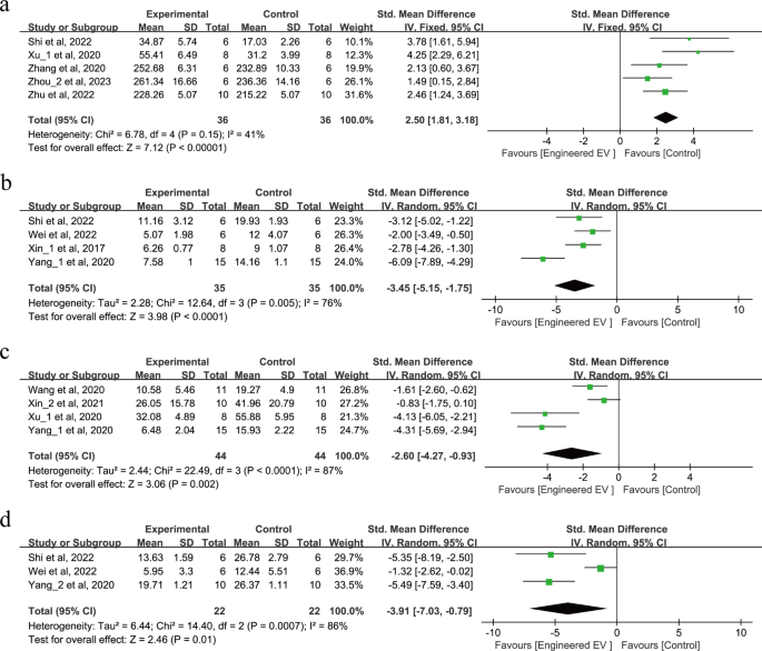
Behavioral tests were conducted on a total of 274 animals across 11 studies as shown in Fig. 3a-d. For motor and coordination function, 5 studies performed the rotarod test (SMD = 2.50, 95% CI: 1.81, 3.18, p < 0.00001, I2 = 41%) as shown in Fig. 3a, while 4 studies performed the grid-walking test (SMD = -3.45, 95% CI: -5.15, -1.75, p < 0.0001, Tau2 = 2.28, I2 = 76%) as shown in Fig. 3b. For motor and sensory function, 4 studies performed adhesive removal test (SMD = -2.60, 95% CI: -4.27, -0.93, p = 0.002, Tau2 = 2.44, I2 = 87%) as shown in Fig. 3c. For learning and memory function, 3 studies performed the morris water maze test (SMD = -3.91, 95% CI: -7.03, -0.79, p = 0.01, Tau2 = 6.44, I2 = 86%) as shown in Fig. 3d. In summary, all these tests suggest that treatment with EEVs improves behavioral recovery after IS.
EEVs reduce the release of pro-inflammatory factors after IS
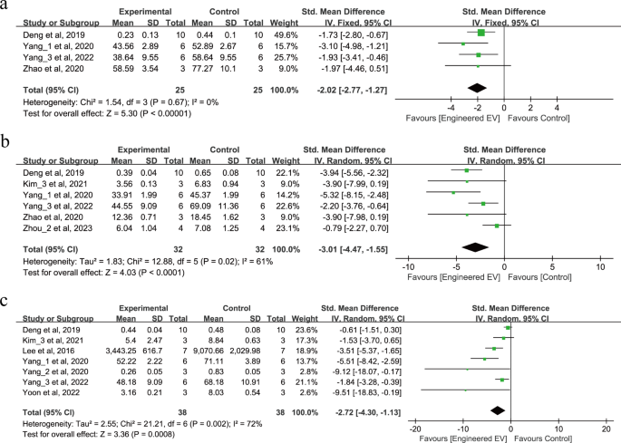
9 studies involving 190 animals reported the release of pro-inflammatory factors after IS as shown in Fig. 4a-c. 4 studies reported that EEVs can reduce IL-1β (SMD = -2.02, 95% CI: -2.77, -1.27, p < 0.00001, I2 = 0%) as shown in Fig. 4a. 6 studies reported that EEVs can reduce the release of IL-6 (SMD = -3.01, 95% CI: -4.47, -1.55, p < 0.0001, Tau2 = 1.83, I2 = 61%) as shown in Fig. 4b. 7 studies reported that EEVs can also reduce the release of TNF-α (SMD = -2.72, 95% CI: -4.30, -1.13, p = 0.0008, Tau2 = 2.55, I2 = 72%) as shown in Fig. 4c. In summary, these studies all demonstrate that treatment with EEVs can reduce the release of pro-inflammatory factors after IS.
EEVs reduce apoptosis rate and increase the number of neurons after IS
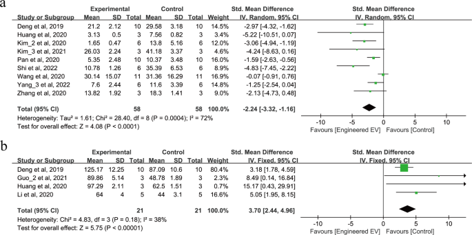
11 studies involving 158 animals reported on the apoptosis rate and the number of neurons after IS, as shown in Fig. 5a-b. 9 studies reported that treatment with EEVs reduce apoptosis rate (SMD = -2.24, 95% CI: -3.32, -1.16, p < 0.0001, Tau2 = 1.61, I2 = 72%) as shown in Fig. 5a. 4 studies reported that treatment with EEVs significantly increase neuron numbers after IS (SMD = 3.70, 95% CI: 2.44, 4.96, p < 0.00001, I2 = 38%) as shown in Fig. 5b.
Subgroup and sensitivity analyses
We conducted a subgroup analysis to explore the source of heterogeneity. As shown in Table 2, we did not observe significant sources of heterogeneity in the outcome of infarct volume among subgroups of randomization, blinding, animal species, source of EVs, methods of engineering, engineering targets, route of administration, and the timepoint of administration. We also conducted a sensitivity analysis to ensure the robustness of determining the overall effect size of the observed outcome measurements. We removed one study at a time and recalculated the pooled effect size for the remaining studies. The results showed that for all outcomes, there was no significant improvement in heterogeneity between studies, indicating that no study had driven the source of heterogeneity.
Research quality and bias risk
As shown in Table 3, the median quality assessment score for the studies was 7 points (IQR: 6–9). However, most studies employed the principle of random allocation and only a few reported concealment of allocation. Half of the studies used a blinding to evaluate the results. Only one study provided information on sample size calculation, which received a risk of bias score of 10 points, as shown in Table 4.
Publication bias
We also conducted a publication bias test and generated funnel plots for outcome measures that included more than ten studies. The results indicated publication bias for both of our outcome measures. The funnel plots for infarct volume and neurological scores appeared asymmetrical, as illustrated in Fig. 6, with a majority of the studies indicating more positive effects of EEVs.
Discussion
Our meta-analysis of 28 published preclinical studies examined the therapeutic effects of EEVs on IS. Our findings showed that treatment with EEVs significantly reduced infarct volume, improved neurological function, and promoted behavioral recovery compared to treatment with native EVs. The observed benefits of EEVs may be attributed to their ability to inhibit apoptosis, increase the number of neurons, and reduce the release of pro-inflammatory factors, as shown in Fig. 7.
Research bias and heterogeneity
As with any meta-analysis, it is essential to consider the potential risks of bias and heterogeneity in the included studies. We assessed the risk of bias using the CAMARADES checklist and found that most studies had a low risk of bias. The median quality assessment score was 7 (IQR: 6–9). As most trials employed random grouping, we did not observe detailed calculations for allocation concealment and sample size. Regarding heterogeneity, we observed moderate to high levels of heterogeneity across the studies, which may be due to differences in sources of EVs, engineered methods, and treatment methods. Despite these limitations, the consistent direction and magnitude of the effect across the studies support the overall conclusions of our meta-analysis.
The potential therapeutic mechanism of EEVs
The potential therapeutic mechanism of EEVs in the treatment of IS is thought to be similar to that of natural EVs. Studies suggest that EVs improve the immune microenvironment at IS sites, inhibit inflammatory reactions, counter cell apoptosis, and promote vascular and nerve remodeling [48,49,50]. However, EEVs may have greater therapeutic potential due to their ability to be engineered to target specific cells or pathways. For example, a study by Yang et al. [40] found that EEVs loaded with circular RNA SCMH1 (circSCMH1) were able to reduce the inhibition of methylCpG-binding protein 2 (MeCP2) target gene transcription, leading to enhanced neuronal plasticity, inhibition of glial cell activation, and improved functional recovery in rats and monkeys after stroke. Another study by Deng et al. [21] found that upregulation of miR-138-5p in EEVs derived from bone marrow mesenchymal stem cells successfully targeted LCN2, promoting astrocyte proliferation, inhibiting inflammatory reactions, inhibiting cell apoptosis, and reducing nerve injury. Similarly, Pan et al. [32] found that EVs rich in miR-132-3p may reduce the production of reactive oxygen species (ROS), BBB dysfunction and brain injury in vascular endothelial cells injured by hypoxia/reperfusion (H/R) by activating PI3K and eNOS. Although EEVs appear promising in targeting specific cellular functions, the research in this domain remains limited, further research is necessary to comprehensively grasp the therapeutic potential of EEVs in IS treatment. Before EEVs can be extensively adopted for IS treatment, conducting additional clinical trials to ascertain their safety and efficacy is imperative.
Preparation of EEVs
There are two strategies for preparing EEVs [8]. The first involves engineering the parent cells, such as pre-treating or transfecting with specific molecules. The second strategy is to directly engineer the EVs with precision, using techniques like electroporation, co-incubation, antibody-specific loading, heat shock or freeze-thaw methods, and ultrasonic treatment. These strategies have been successfully implemented in the field of EEVs. The subgroup analysis in our study explored the differences in the effects of these two strategies on reducing infarct volume. Approximately half of the studies involved engineering the parent cells, and our observations suggest that directly engineering the EVs might be more effective in reducing infarct volume compared to engineering the parent cells. However, this difference was not statistically significant and further research is needed for validation. Utilizing parent cells to produce EEVs offers certain advantages: it is simpler and more convenient, and it retains the biophysical characteristics and stability of the EVs. However, this method also carries inherent risks. Overexpression of certain molecules in cells can trigger complex biological reactions, possibly compromising the bioactivity of EVs [8]. In contrast, direct engineering of EVs may provide greater control, ensuring precision in loading, targeting, and delivering the functions of EVs. Current engineering modifications to EVs mainly focus on enhancing their loading capacity, circulation time, and targeting abilities to achieve better therapeutic outcomes.
Loading capacity of EEVs
The ability to deliver various substances to specific cells makes EVs promising candidates for drug delivery. Research indicates that enhancing the miRNA content in EVs can potentially ameliorate ischemic brain injury [34, 37, 47]. For example, Wang et al. [34] found that EEVs rich in miR-126 are more effective than natural EVs in treating diabetes-induced ischemia by reducing acute injury and promoting neural recovery. Similarly, Xin et al. [37] showed that secretion rich in miR-17-92 increased axonal elongation and myelin formation in rats by down-regulating the PTEN-induced PI3K/Akt/mTOR pathway, thus aiding in nerve function recovery post-middle cerebral artery occlusion. In addition, EVs have been explored as drug delivery vehicles. Engineering technologies can facilitate the loading of commonly used drugs for treating ischemic injuries into EVs. Such approaches can reduce the damage and inactivation of drugs during transportation, improving their bioavailability and specificity. For example, Zhu et al. [47] showed that EEVs loaded with brain-derived neurotrophic factor (BDNF) not only inhibited the activation of microglia after stroke but also promoted the differentiation of endogenous neural stem cells into neurons. Guo et al. [23] found that EEVs loaded with quercetin (Que) can activate Nrf2/HO-1 pathway to inhibit ROS production and improve the survival rate of neurons.
Circulation time and targeting of EVs
In recent years, researchers have increasingly focused on enhancing the stability, circulation half-life, and targeting capabilities of EVs within the body. Although previous studies have shown that EVs are often rapidly cleared or concentrated in the liver, spleen, and lungs after injection into animal models, the therapeutic effect of EVs is closely related to their half-life and effective concentration in the lesion area [51, 52]. To extend the circulation half-life, scientists have begun to use nanotechnology to encapsulate EVs, thereby reducing their non-specific interactions with other cells, and subsequently enhancing the stability and half-life of EVs [53, 54]. For instance, Liu et al. [54] used hyaluronic acid hydrogel to encapsulate exosomes derived from bone marrow mesenchymal stem cells, thus achieving higher stability and promoting brain structure reconstruction and neurological function recovery. Additionally, to enhance the targeting capabilities of EVs, researchers have started to engineer the EVs. For example, Tian et al. [55] coupled c(RGDyK) peptide to the surface of EVs, enabling the intravenously injected cRGD-Exo to specifically target ischemic brain lesion areas. Alvarez et al. [56] enhanced targeting to neurons by engineering dendritic cells to express Lamp2b (an EV membrane protein) fused with neuron-specific RVG peptide. Kim et al. [27] also developed magnetic EVs fused with iron oxide nanoparticle (IONP) as a bioengineering means to enhance the targeting ability of EVs. In summary, through nanotechnology and engineering approaches, researchers are actively exploring ways to prolong the circulation half-life and enhance the targeting capability of EVs to optimize their therapeutic applications.
Limitations
Our review has some limitations. First, only a few studies were included. Significant differences exist in the following aspects of the covered studies: source, injection route, treatment time, dose, and follow-up time of EVs in the covered studies. Additionally, there are concerns about data deviation and accuracy which require further evaluation. Future research should be more comprehensive to address these limitations.
Conclusions
Our study highlights the advantages of EEVs in the treatment of IS. Compared to natural EVs, they have shown a stronger therapeutic effect, especially in reducing infarct volume, enhancing neural function, promoting behavioral recovery, reducing inflammatory responses, regulating cell apoptosis, and increasing the number of neurons. These findings open a new perspective for the in-depth study of EV engineering techniques. However, the production and optimization of EEVs do face challenges in terms of time and cost. Although this field remains in its exploratory phase, further investigations are imperative to deepen our grasp of the therapeutic potential of EEVs, particularly in the context of IS treatment. Before the clinical introduction of EEVs, it is crucial to conduct further clinical trials to ensure both their safety and efficacy.
Data Availability
All the data that underlie the conclusions in this research are available online.
Abbreviations
- ADSC:
-
Adipose-derived stromal cell
- ANG1:
-
Angiopoietin 1
- ANG2:
-
Angiopoietin 2
- BBB:
-
Blood-brain barrier
- BDNF:
-
Brain-derived neurotrophic factor
- BMSC:
-
Bone marrow mesenchymal stem cell
- CAMARADES:
-
Collaborative Approach to Meta-analysis and Review of Animal Data in Experimental Studies
- EEVs:
-
Engineered extracellular vesicles
- EVs:
-
Extracellular vesicles
- FDA:
-
Food and Drug Administration
- IS:
-
Ischemic stroke
- MCAO:
-
Middle cerebral artery occlusion
- MMP9:
-
Matrix metallopeptidase 9
- mNSS:
-
Modified neurological severity score
- MVs:
-
Microvesicles
- NSC:
-
Neural stem cell
- PRISMA:
-
Preferred Reporting Items for Systematic Reviews and Meta-Analyses
- ROS:
-
Reactive oxygen species
- TSP1:
-
Thrombopoietin 1
- VEGF:
-
Vascular endothelial growth factor
References
Donnan GA, Fisher M, Macleod M, et al. Stroke Lancet. 2008;371:1612–23. https://doi.org/10.1016/S0140-6736(08)60694-7
Marei HE, Hasan A, Rizzi R, et al. Potential of stem cell-based therapy for ischemic stroke. Front Neurol. 2018;9:34. https://doi.org/10.3389/fneur.2018.00034
Ghosh D, Sehgal K, Sodnar B, et al. Drug repurposing for stroke intervention. Drug Discovery Today. 2022;27(7):1974–82. https://doi.org/10.1016/j.drudis.2022.03.003
Suzuki K, Matsumaru Y, Takeuchi M, et al. Effect of mechanical thrombectomy without vs with intravenous thrombolysis on functional outcome among patients with acute ischemic stroke: the skip randomized clinical trial. JAMA. 2021;325(3):244–53. https://doi.org/10.1001/jama.2020.23522
Zi W, Qiu Z, Li F, et al. Effect of endovascular treatment alone vs intravenous alteplase plus endovascular treatment on functional independence in patients with acute ischemic stroke: the devt randomized clinical trial. JAMA. 2021;325(3):234–43. https://doi.org/10.1001/jama.2020.23523
Shtam TA, Kovalev RA, Varfolomeeva EY, et al. Exosomes are natural carriers of exogenous siRNA to human cells in vitro. Cell Commun Signal. 2013;11:88. https://doi.org/10.1186/1478-811X-11-88
Budnik V, Ruiz-Cañada C, Wendler F. Extracellular vesicles round off communication in the nervous system. Nat Rev Neurosci. 2016;17(3):160–72. https://doi.org/10.1038/nrn.2015.29
De Abreu RC, Fernandes H, da Costa Martins PA, et al. Native and bioengineered extracellular vesicles for cardiovascular therapeutics. Nat Rev Cardiol. 2020;17(11):685–97. https://doi.org/10.1038/s41569-020-0389-5
Ferrantelli F, Chiozzini C, Leone P, et al. Engineered Extracellular Vesicles/Exosomes as a New Tool against Neurodegenerative Diseases. Pharmaceutics. 2020;12(6):529. https://doi.org/10.3390/pharmaceutics12060529
Rufino-Ramos D, Albuquerque PR, Carmona V, et al. Extracellular vesicles: novel promising delivery systems for therapy of brain diseases. J Control Release. 2017;262:247–58. https://doi.org/10.1016/j.jconrel.2017.07.001
Gratpain V, Mwema A, Labrak Y, et al. Extracellular vesicles for the treatment of central nervous system diseases. Adv Drug Deliv Rev. 2021;174:535–52. https://doi.org/10.1016/j.addr.2021.05.006
Khan H, Pan JJ, Li Y, et al. Native and Bioengineered Exosomes for ischemic stroke therapy. Front Cell Dev Biol. 2021;9:619565. https://doi.org/10.3389/fcell.2021.619565
Cun Y, Jin Y, Wu D, et al. Exosome in crosstalk between inflammation and angiogenesis: a potential therapeutic strategy for stroke. Mediators Inflamm. 2022;2022:7006281. https://doi.org/10.1155/2022/7006281
Tian T, Cao L, He C, et al. Targeted delivery of neural progenitor cell-derived extracellular vesicles for anti-inflammation after cerebral ischemia. Theranostics. 2021;11(13):6507–21. https://doi.org/10.7150/thno.56367
Zhang Y, Liu J, Su M, et al. Exosomal microRNA-22-3p alleviates cerebral ischemic injury by modulating KDM6B/BMP2/BMF axis. Stem Cell Res Ther. 2021;12(1):111. https://doi.org/10.1186/s13287-020-02091-x
Mathiyalagan P, Liang Y, Kim D, et al. Angiogenic mechanisms of human CD34 + stem cell exosomes in the repair of ischemic hindlimb. Circ Res. 2017;120(9):1466–76.
Xu X, Zhang H, Li J, et al. Combination of EPC-EXs and NPC-EXs with miR-126 and miR-210 overexpression produces better therapeutic effects on ischemic stroke by protecting neurons through the Nox2/ROS and BDNF/TrkB pathways. Exp Neurol. 2023;359:114235. https://doi.org/10.1016/j.expneurol.2022.114235
Villa A, Garofalo M, Crescenti D, et al. Transplantation of autologous extracellular vesicles for cancer-specific targeting. Theranostics. 2021;11(5):2034–47. https://doi.org/10.7150/thno.51344
Moher D, Liberati A, Tetzlaff J, et al. Preferred reporting items for systematic reviews and meta-analyses: the PRISMA statement. PLoS Med. 2009;6(7):e1000097. https://doi.org/10.1371/journal.pmed.1000097
Macleod MR, O’Collins T, Howells DW, et al. Pooling of animal experimental data reveals influence of study design and publication bias. Stroke. 2004;35(5):1203–8. https://doi.org/10.1161/01.STR.0000125719.25853.20
Deng Y, Chen D, Gao F, et al. Exosomes derived from microRNA-138-5p-overexpressing bone marrow-derived mesenchymal stem cells confer neuroprotection to astrocytes following ischemic stroke via inhibition of LCN2. J Biol Eng. 2019;13:71. https://doi.org/10.1186/s13036-019-0193-0
Guo L, Pan J, Li F, et al. A novel brain targeted plasma exosomes enhance the neuroprotective efficacy of edaravone in ischemic stroke. IET Nanobiotechnol. 2021;15(1):107–16. https://doi.org/10.1049/nbt2.12003
Guo L, Huang Z, Huang L, et al. Surface-modified engineered exosomes attenuated cerebral ischemia/reperfusion injury by targeting the delivery of quercetin towards impaired neurons. J Nanobiotechnol. 2021;19(1):141. https://doi.org/10.1186/s12951-021-00879-4
Huang Z, Guo L, Huang L, et al. Baicalin-loaded macrophage-derived exosomes ameliorate ischemic brain injury via the antioxidative pathway. Mater Sci Eng C Mater Biol Appl. 2021;126:112123. https://doi.org/10.1016/j.msec.2021.112123
Jiang Y, Wang R, Wang C, et al. Brain microenvironment responsive and pro-angiogenic extracellular vesicle-hydrogel for promoting Neurobehavioral Recovery in type 2 Diabetic mice after stroke. Adv Healthc Mater. 2022;11(22):e2201150. https://doi.org/10.1002/adhm.202201150
Kim M, Kim G, Hwang DW, et al. Delivery of high mobility Group Box-1 siRNA using brain-targeting exosomes for ischemic stroke therapy. J Biomed Nanotechnol. 2019;15(12):2401–12. https://doi.org/10.1166/jbn.2019.2866
Kim HY, Kim TJ, Kang L, et al. Mesenchymal stem cell-derived magnetic extracellular nanovesicles for targeting and treatment of ischemic stroke. Biomaterials. 2020;243:119942. https://doi.org/10.1016/j.biomaterials.2020.119942
Kim M, Lee Y, Lee M. Hypoxia-specific anti-RAGE exosomes for nose-to-brain delivery of anti-miR-181a oligonucleotide in an ischemic stroke model. Nanoscale. 2021;13(33):14166–78. https://doi.org/10.1039/d0nr07516g
Lee JY, Kim E, Choi SM, et al. Microvesicles from brain-extract-treated mesenchymal stem cells improve neurological functions in a rat model of ischemic stroke. Sci Rep. 2016;6:33038. https://doi.org/10.1038/srep33038
Li F, Zhao L, Shi Y, et al. Edaravone-Loaded macrophage-derived Exosomes Enhance Neuroprotection in the Rat Permanent Middle cerebral artery occlusion model of stroke. Mol Pharm. 2020;17(9):3192–201. https://doi.org/10.1021/acs.molpharmaceut.0c00245
Liu W, Su C, Qi Y, et al. Brain-targeted heptapeptide-loaded exosomes attenuated ischemia-reperfusion injury by promoting the transfer of healthy mitochondria from astrocytes to neurons. J Nanobiotechnol. 2022;20(1):242. https://doi.org/10.1186/s12951-022-01425-6
Pan Q, Kuang X, Cai S, et al. Mir-132-3p priming enhances the effects of mesenchymal stromal cell-derived exosomes on ameliorating brain ischemic injury. Stem Cell Res Ther. 2020;11(1):260. https://doi.org/10.1186/s13287-020-01761-0
Shi X, Zhong X, Deng L, et al. Mesenchymal stem cell-derived extracellular vesicle-enclosed microRNA-93 prevents hypoxic-ischemic brain damage in rats. Neuroscience. 2022;500:12–25. https://doi.org/10.1016/j.neuroscience.2022.06.037
Wang J, Chen S, Zhang W, et al. Exosomes from miRNA-126-modified endothelial progenitor cells alleviate brain injury and promote functional recovery after stroke. CNS Neurosci Ther. 2020;26(12):1255–65. https://doi.org/10.1111/cns.13455
Wei R, Zhang L, Hu W, et al. Zeb2/Axin2-Enriched BMSC-Derived Exosomes promote post-stroke functional recovery by enhancing neurogenesis and neural plasticity. J Mol Neurosci. 2022;72(1):69–81. https://doi.org/10.1007/s12031-021-01887-7
Xin H, Katakowski M, Wang F, et al. MicroRNA cluster mir-17-92 cluster in Exosomes Enhance Neuroplasticity and functional recovery after stroke in rats. Stroke. 2017;48(3):747–53. https://doi.org/10.1161/STROKEAHA.116.015204
Xin H, Liu Z, Buller B, et al. MiR-17-92 enriched exosomes derived from multipotent mesenchymal stromal cells enhance axon-myelin remodeling and motor electrophysiological recovery after stroke. J Cereb Blood Flow Metab. 2021;41(5):1131–44. https://doi.org/10.1177/0271678X20950489
Xu L, Ji H, Jiang Y, et al. Exosomes Derived from CircAkap7-Modified adipose-derived mesenchymal stem cells protect against cerebral ischemic Injury. Front Cell Dev Biol. 2020;8:569977. https://doi.org/10.3389/fcell.2020.569977
Yang HC, Zhang M, Wu R, et al. C-C chemokine receptor type 2-overexpressing exosomes alleviated experimental post-stroke cognitive impairment by enhancing microglia/macrophage M2 polarization. World J Stem Cells. 2020;12(2):152–67. https://doi.org/10.4252/wjsc.v12.i2.152
Yang L, Han B, Zhang Z, et al. Extracellular vesicle-mediated delivery of circular RNA SCMH1 promotes functional recovery in Rodent and Nonhuman Primate Ischemic Stroke Models. Circulation. 2020;142(6):556–74. https://doi.org/10.1161/CIRCULATIONAHA.120.045765
Yang H, Chen J. Bone marrow mesenchymal stem cell-derived exosomes carrying long noncoding RNA ZFAS1 alleviate oxidative stress and inflammation in ischemic stroke by inhibiting microRNA-15a-5p. Metab Brain Dis. 2022;37(7):2545–57. https://doi.org/10.1007/s11011-022-00997-4
Yoon EJ, Choi Y, Kim TM, et al. The neuroprotective Effects of Exosomes Derived from TSG101-Overexpressing human neural stem cells in a stroke model. Int J Mol Sci. 2022;23(17):9532. https://doi.org/10.3390/ijms23179532
Zhang G, Zhu Z, Wang H, et al. Exosomes derived from human neural stem cells stimulated by interferon gamma improve therapeutic ability in ischemic stroke model. J Adv Res. 2020;24:435–45. https://doi.org/10.1016/j.jare.2020.05.017
Zhao Y, Gan Y, Xu G, et al. Exosomes from MSCs overexpressing microRNA-223-3p attenuate cerebral ischemia through inhibiting microglial M1 polarization mediated inflammation. Life Sci. 2020;260:118403. https://doi.org/10.1016/j.lfs.2020.118403
Zhou X, Deng X, Liu M, et al. Intranasal delivery of BDNF-loaded small extracellular vesicles for cerebral ischemia therapy. J Control Release. 2023;357:1–19. https://doi.org/10.1016/j.jconrel.2023.03.033. Epub 2023 Mar 28. PMID: 36958402.
Zhou H, Zhou J, Teng H, Yang H, Qiu J, Li X. MiR-145 enriched exosomes derived from bone marrow-derived mesenchymal stem cells protects against cerebral ischemia-reperfusion injury through downregulation of FOXO1. Biochem Biophys Res Commun. 2022;632:92–9. https://doi.org/10.1016/j.bbrc.2022.09.089
Zhu ZH, Jia F, Ahmed W, et al. Neural stem cell-derived exosome as a nano-sized carrier for BDNF delivery to a rat model of ischemic stroke. Neural Regen Res. 2023;18(2):404–9. https://doi.org/10.4103/1673-5374.346466
Spellicy SE, Stice SL. Tissue and stem cell sourced Extracellular Vesicle Communications with Microglia. Stem Cell Rev Rep. 2021;17(2):357–68. https://doi.org/10.1007/s12015-020-10011-y
Tariq MB, Lee J, McCullough LD. Sex differences in the inflammatory response to stroke. Semin Immunopathol. 2022. https://doi.org/10.1007/s00281-022-00969-x
Yang C, Yuan F, Shao W, et al. Protective role of exosomes derived from regulatory T cells against inflammation and apoptosis of BV-2 microglia under oxygen-glucose deprivation/reperfusion challenge. Genet Mol Biol. 2022;45(4):e20220119. https://doi.org/10.1590/1678-4685-GMB-2022-0119
Aimaletdinov AM, Gomzikova MO. Tracking of Extracellular vesicles’ biodistribution: New Methods and Approaches. Int J Mol Sci. 2022;23(19):11312. https://doi.org/10.3390/ijms231911312
Toh WS, Zhang B, Lai RC, et al. Immune regulatory targets of mesenchymal stromal cell exosomes/small extracellular vesicles in tissue regeneration. Cytotherapy. 2018;20(12):1419–26. https://doi.org/10.1016/j.jcyt.2018.09.008
Emam SE, Elsadek NE, Abu Lila AS, et al. Anti-PEG IgM production and accelerated blood clearance phenomenon after the administration of PEGylated exosomes in mice. J Control Release. 2021;334:327–34. https://doi.org/10.1016/j.jconrel.2021.05.001
Liu X, Wu C, Zhang Y, et al. Hyaluronan-based hydrogel integrating exosomes for traumatic brain injury repair by promoting angiogenesis and neurogenesis. Carbohydr Polym. 2023;306:120578. https://doi.org/10.1016/j.carbpol.2023.120578
Tian T, Zhang HX, He CP, et al. Surface functionalized exosomes as targeted drug delivery vehicles for cerebral ischemia therapy. Biomaterials. 2018;150:137–49. https://doi.org/10.1016/j.biomaterials
Alvarez-Erviti L, Seow Y, Yin H, et al. Delivery of siRNA to the mouse brain by systemic injection of targeted exosomes. Nat Biotechnol. 2011;29(4):341–5. https://doi.org/10.1038/nbt.1807
Acknowledgements
Not applicable.
Funding
This research was supported by the National High Level Hospital Clinical Research Funding (2022-PUMCH-C-032), the Beijing Tianjin Hebei basic research cooperation project (19JCZDJC64600(Z)), and the National Key R&D Program of China (2018YFA0108603) for research design, data collection, analysis, and publication.
Author information
Authors and Affiliations
Contributions
Research concept and design: PTL, RY; Data acquisition: RY, SHW, QH, YHC, JBC, HSX; Data analysis and interpretation: PTL, XYL, LY, XZ; Draft: PTL; Revised version: JJW,QH; Source of funds: JJW. PTL and RY have the same contribution to this research. All authors have read and approved the manuscript.
Corresponding authors
Ethics declarations
Ethics approval and consent to participate
The publication of this article has been approved by all authors.
Consent for publication
Not applicable.
Conflict of interest
All authors confirm that there is no conflict of interest in this research.
Additional information
Publisher’s Note
Springer Nature remains neutral with regard to jurisdictional claims in published maps and institutional affiliations.
Electronic supplementary material
Below is the link to the electronic supplementary material.
Rights and permissions
Open Access This article is licensed under a Creative Commons Attribution 4.0 International License, which permits use, sharing, adaptation, distribution and reproduction in any medium or format, as long as you give appropriate credit to the original author(s) and the source, provide a link to the Creative Commons licence, and indicate if changes were made. The images or other third party material in this article are included in the article’s Creative Commons licence, unless indicated otherwise in a credit line to the material. If material is not included in the article’s Creative Commons licence and your intended use is not permitted by statutory regulation or exceeds the permitted use, you will need to obtain permission directly from the copyright holder. To view a copy of this licence, visit http://creativecommons.org/licenses/by/4.0/. The Creative Commons Public Domain Dedication waiver (http://creativecommons.org/publicdomain/zero/1.0/) applies to the data made available in this article, unless otherwise stated in a credit line to the data.
About this article
Cite this article
Li, P., Yin, R., Chen, Y. et al. Engineered extracellular vesicles for ischemic stroke: a systematic review and meta-analysis of preclinical studies. J Nanobiotechnol 21, 396 (2023). https://doi.org/10.1186/s12951-023-02114-8
Received:
Accepted:
Published:
DOI: https://doi.org/10.1186/s12951-023-02114-8
